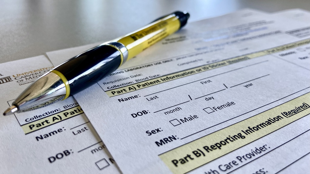
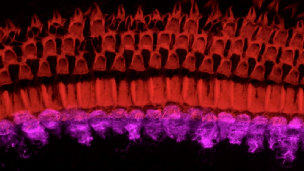
MORL Logo

Making Lives Better

Part of Molecular Otolaryngology and Renal Research Laboratories' requisition form with black and gold pen.

Requisition Form

Light microscopy of a kidney biopsy using a Jones stain and showing the typical "double contour" appearance of the glomerular capillary walls, indicating the presence of dense deposits that stain poorly with silver.

Complement-Mediated Kidney Disease Division

Confocal microscope image of a whole mount preparation of a mouse cochlea showing the cochlear hair cells. The inner hair cells (bottom) are responsible for the mechano-transduction of sound into an electrical signal and are stained in magenta (Vglut3), while the three rows of outer hair cells are labeled in red (phalloidin).  Note that some of the outer hair cells in the outermost row (top) are missing.

Hearing Loss Division